Specialties
Cardiology
- Cardiac catheterization
- Electrocardiographic (EKG) Stress Testing
- Echocardiography
- Electrocardiography
- Electrophysiology
- Women's cardiac care

Cardiac Catheterization
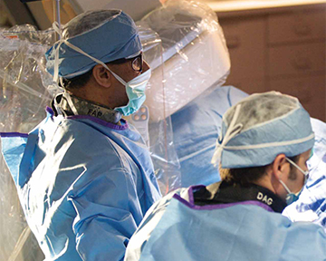
Cardiac catheterization is a procedure used to diagnose and treat conditions of the heart with the use of a long, thin tube called a catheter. The catheter is inserted into a blood vessel in the arm, groin or neck area and then guided to the heart. At the heart, the catheter can be used to:
- Detect any blockages or abnormalities
- Take a blood or muscle sample
- Measure blood pressure and oxygen levels
- Detect congenital heart defects

Stress Testing (EKG, Nuclear, Pharmacologic)
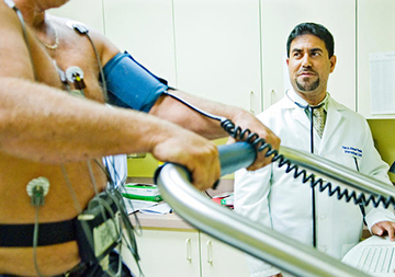
An EKG stress test, also known as an exercise tolerance test or cardiac stress test, evaluates the adequacy of the blood flow to the heart as increasing amounts of exercise are performed in a closely monitored setting. Normally, the heart rate and blood pressure increase during exercise, and the electrocardiogram (EKG: a recording of the electrical activity of the heart) should show little change, except for the increase in heart rate. However, in the presence of certain kinds of heart conditions, the stress of exercise can cause abnormal changes in the heart rhythm, blood pressure, or EKG. In this way, the stress test can help the doctor assess for the presence of some types of heart disease.
Echocardiography
An echocardiogram (also called an echo) is a type of ultrasound test that uses high-pitched sound waves that are sent through a device called a transducer. The device picks up echoes of the sound waves as they bounce off the different parts of your heart. These echoes are turned into moving pictures of your heart that can be seen on a video screen. The cardiologist will look at the images and generate a report to your physician.
The different types of echocardiograms are:
Transthoracic echocardiogram (TTE) – This is the most common type. Views of the heart are obtained by moving the transducer to different locations on your chest or abdominal wall.

This test is done to:
- Look for the causes of abnormal heart sounds (murmurs or clicks), an enlarged heart, unexplained chest pains, shortness of breath, or irregular heartbeats.
- Check the thickness and movement of the heart wall.
- Look at the heart valves and check how well they work.
- See how well an artificial heart valve is working.
- Measure the size and shape of the heart’s chambers.
- Check the ability of your heart chambers to pump blood (cardiac performance). During an echocardiogram, your doctor can calculate the how much blood your heart is pumping during each heartbeat (ejection fraction). You might have a low ejection fraction if you have heart failure.
- Detect a disease that affects the heart muscle and the way it pumps, such as cardiomyopathy.
At times you will be asked to hold very still, breathe in and out very slowly, hold your breath, or lie on your left side. The transducer is usually moved to different areas on your chest that provides specific views of your heart. The test usually takes 30 to 45 minutes. When the test is over, the gel is wiped off and the electrodes are removed.
Transesophageal echocardiogram (TEE) – A transesophageal echocardiogram (TEE), like the transthoracic echocardiogram, uses sound waves to make pictures of your heart and valves. For this test, the probe is passed down the esophagus instead of being moved over the outside of the chest wall. TEE shows clearer pictures of your heart because the probe is located closer to the heart and because the lungs and bones of the chest wall do not block the sound waves produced by the probe.
An electrocardiogram (also called EKG or ECG) is a test that records the electrical activity of your heart through small electrode patches attached to the skin of your chest, arms, and legs. An EKG may be part of a routine physical exam or it may be used as a test for heart disease. EKGs are quick, safe and painless and are routinely performed if a heart condition is suspected. An EKG can be used to further investigate symptoms related to heart problems.
Your doctor uses the EKG to:
- Assess your heart rhythm.
- Diagnose poor blood flow to the heart muscle (ischemia).
- Diagnose a heart attack.
- Evaluate certain abnormalities of your heart, such as an enlarged heart.
Electrophysiology testing makes it possible to study heart rhythm disturbances under controlled circumstances. By using special insulated wires called catheters, the doctor is able to identify rhythm disturbance and choose the best method of treatment. Intravenous catheters are placed, usually from the vein in the groin area, so that they can be advanced into the right side of the heart. In the right side of the heart, recordings can be made that give a very clear picture of the normal sequence of electrical activation within the heart.
In addition, the heart can be submitted in a variety of ways in an attempt to elicit abnormal rhythm if they exist. After the catheters are in position, the doctor will evaluates the heart rhythm disturbance by giving the heart small electrical impulses (by artificial pacemaker through one of the catheters) to make it beat at a different rates. This offers important information regarding arrhythmia mechanism, prognosis, risk of serious symptoms and response to medications.
Holter Monitor
A Holter Monitor is a battery operated device that records a 24 or 48 hour electrocardiogram (EKG). An EKG records the electrical activity of your heart. Five electrodes (“sticky patches”) will be attached to your chest. These electrodes are attached to the recorder, which is about the size and weight of a transistor radio or “walkman”.
The Holter Monitor is worn for 24 or 48 hours (depending on the physician’s order). It will be attached to a strap, so it will be easier for you to carry around with you. You will be given a small diary to keep notes in. If you have any symptoms such as dizziness, chest pain, difficulty breathing or feelings that your heart is skipping a beat, please write the time they occur in your diary.
During the 24 or 48 hours that you have the Holter Monitor on: DO NOT shower. Keep the monitor on while you sleep. If you have any problems with the sticky patches or the recorder, let your health care provider know.
Upon completion of the monitoring period, the electrodes (“sticky patches”) will be removed. The monitor is to be returned to the office in order to retrieve the information recorded. The tape and the diary will be reviewed and interpreted by a Cardiology physician and a report will be sent to your referring provider.
Nuclear Cardiology
A nuclear stress test, also referred to as a myocardial perfusion scan, is one of the most commonly performed diagnostic heart tests. This technology makes use of small amounts of radioactive tracers to assess blood flow to the heart muscle and accurately detect the presence and extent of obstructive coronary artery disease. Dual-isotope stress testing is performed in conjunction with treadmill exercise or a special medication for patients who are not able to exercise. Three-dimensional, computer-generated images through the heart are created, allowing the doctor to see blood flow abnormalities in a multi-view color display. Important data regarding the size and function of the heart’s pumping chambers are also provided by these images.
A Nuclear Cardiology Stress Test is also known as a Thallium Stress Test, a Dual Isotope Test, or a Myocardial Perfusion Imaging Stress Test. It is usually performed in nuclear medicine laboratories. During the test you will be given two injections of radioactive contrast materials to determine if your heart is receiving adequate blood flow during rest and stress conditions.
Some of the radioactive injections (also called isotopes) that may be used are:
- Myoview (99m Technetium Tetrofosmin)
- Cardiolite (99m Technetium Sestamibi)
At Padder Health, we provide specialized prevention, diagnosis and treatment services for women at risk or with a history for cardiovascular conditions such as heart disease, atherosclerotic disease, arrhythmia and heart failure. Based on a physical examination and evaluation of the patient’s medical history, family history and lifestyle, an individualized treatment approach will be developed to promote lifelong heart health through simple modifications.
Screening for heart disease is recommended for women over the age of 18 who have one or more risk factors of a cardiovascular condition, including obesity, diabetes, high blood pressure, smoking or a family history of heart disease. We also provide educational services to these women to help them prevent heart disease and maintain an active, healthy lifestyle.