-
 August 29, 2026
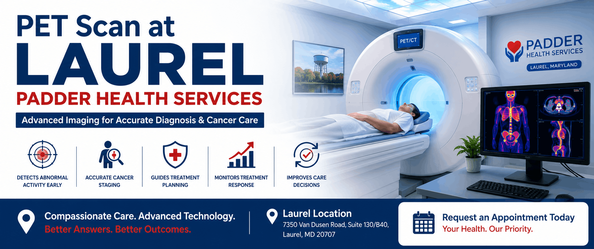
When doctors need more information about what is happening inside the body, advanced medical imaging can provide important clues. A PET scan, or positron emission tomography scan, is a type of nuclear medicine imaging that shows areas of activity inside the body.
PET imaging is particularly important in cancer care because many cancer cells use glucose differently from normal cells. A commonly used PET tracer, called FDG, is a radioactive form of glucose. Areas that take up more of the tracer can appear differently on PET images and may help physicians identify areas that require further evaluation. ([cancer.gov](https://www.cancer.gov/about-cancer/diagnosis-staging/ct-scans-fact-sheet?utm_source=chatgpt.com))
When PET is combined with CT, the resulting PET/CT scan provides both functional information and detailed anatomical information. This combination can help physicians evaluate disease, determine the extent of cancer, plan treatment and assess how well treatment is working. ([cancer.gov](https://www.cancer.gov/publications/dictionaries/cancer-terms/def/pet-ct-scan?utm_source=chatgpt.com))
What Is a PET Scan?
A PET scan is an imaging examination that uses a small amount of a radioactive tracer to show metabolic or functional activity in tissues.
Unlike imaging that primarily shows anatomy, PET can provide information about how tissues are functioning. When combined with CT, physicians can compare areas of increased or decreased activity with the body’s anatomical structures.
🚨 PET Scans Are Not Emergency Tests
A PET scan is generally a planned diagnostic examination. If you are experiencing severe chest pain, severe difficulty breathing, sudden weakness, loss of consciousness or another potentially life-threatening symptom, seek emergency medical care immediately rather than waiting for a scheduled imaging appointment.
What Is a PET/CT Scan?
A PET/CT scan combines two imaging technologies in one examination. The PET portion provides functional information about activity within tissues, while CT provides detailed anatomical images showing the location and structure of organs and tissues.
The combined information can provide a more complete picture than either examination alone. PET/CT is used in a variety of clinical situations, particularly in oncology, where understanding both the location and activity of abnormal tissue can be important. ([cancer.gov](https://www.cancer.gov/about-cancer/diagnosis-staging/ct-scans-fact-sheet?utm_source=chatgpt.com))
PET Imaging
Shows functional or metabolic activity within tissues.
CT Imaging
Provides detailed anatomical information about organs and structures.
Combined PET/CT
Combines functional and anatomical information in one examination.
Clinical Decision Support
Results can provide information that helps physicians plan further evaluation or treatment.

Why Are PET Scans Used in Cancer Care?
PET/CT can play an important role at several stages of cancer care. Depending on the type of cancer and the tracer used, PET imaging may help physicians evaluate a suspicious abnormality, determine whether cancer has spread, plan treatment or assess how a tumor responds to therapy.
PET imaging is not appropriate for every cancer or every patient. The usefulness of a PET scan depends on the type of disease, the clinical question and the tracer being used.
1Detecting Abnormal Activity
One of the important features of PET imaging is its ability to show areas of increased metabolic activity. Because many cancer cells use glucose at a higher rate than some normal tissues, FDG-PET can help identify areas that may require further investigation. ([cancer.gov](https://www.cancer.gov/about-cancer/diagnosis-staging/ct-scans-fact-sheet?utm_source=chatgpt.com))
However, increased tracer uptake does not automatically mean that an area is cancer. Infection, inflammation and other conditions can also cause increased metabolic activity. A PET scan must therefore be interpreted by qualified specialists alongside other clinical and imaging information.
A PET Scan Does Not Automatically Diagnose Cancer
An area of increased activity on a PET scan can have several possible causes. PET findings should be interpreted together with your medical history, examination and other tests. In some situations, a biopsy may be needed to establish a diagnosis.
2Helping Determine Cancer Stage
Cancer staging describes the extent of cancer, including whether it has spread beyond the original tumor. Determining the stage can help physicians choose an appropriate treatment strategy. ([cancer.gov](https://www.cancer.gov/about-cancer/diagnosis-staging/staging?utm_source=chatgpt.com))
PET/CT may provide information about areas of abnormal metabolic activity throughout the body and can sometimes identify disease that is not obvious from anatomical imaging alone.
The exact role of PET/CT in staging depends on the type of cancer. Some cancers are particularly well suited to PET imaging, while others may be better evaluated with CT, MRI or other tests.
3Finding Possible Spread of Cancer
When cancer spreads from its original location to other parts of the body, the process is called metastasis. PET/CT can sometimes help identify areas that may represent metastatic disease.
This information can be valuable because the presence or absence of disease in other parts of the body may affect treatment planning.
Why Whole-Body Information Can Matter
A PET/CT examination may allow physicians to evaluate multiple regions of the body during one imaging session. This can help provide a broader picture of disease activity when whole-body imaging is clinically appropriate.
4Planning Cancer Treatment
PET/CT findings can help physicians understand where active disease may be located and can contribute to treatment planning.
Depending on the cancer type and treatment strategy, PET imaging may provide information that helps physicians decide whether surgery, radiation therapy, systemic treatment or another approach should be considered.
PET findings are interpreted alongside pathology, physical examination, laboratory tests and other imaging studies rather than being used in isolation.
5Monitoring Response to Treatment
PET imaging can sometimes help physicians determine how a tumor responds to treatment by evaluating changes in metabolic activity.
This can be particularly useful because changes in metabolic activity may occur before obvious changes in tumor size are visible on anatomical imaging.
The timing and type of follow-up PET scan depend on the cancer diagnosis and treatment plan. Your oncology team determines whether repeat PET imaging is appropriate.
6Checking for Possible Cancer Recurrence
After treatment, doctors may sometimes use imaging to investigate whether cancer has returned. PET/CT may be appropriate in selected situations when there is clinical concern for recurrent disease.
Whether PET is recommended depends on the original cancer, previous treatment, symptoms, laboratory findings and other imaging results.
7Different PET Tracers Are Used for Different Purposes
FDG is one of the most widely used PET tracers, but it is not the only option. Different radiopharmaceuticals are designed to provide information about specific biological processes.
For example, specialized PET tracers may be used in selected patients for prostate cancer or neuroendocrine tumors. The appropriate tracer depends on the suspected or known condition and the clinical question being evaluated. ([cancer.gov](https://www.cancer.gov/about-cancer/diagnosis-staging/ct-scans-fact-sheet?utm_source=chatgpt.com))
FDG PET
Uses a glucose-based tracer and is widely used for many oncology applications.
Targeted PET Tracers
Some tracers are designed to target specific biological features associated with particular diseases.
Prostate Imaging
Certain specialized tracers can target prostate-specific membrane antigen in appropriate clinical situations.
Other Applications
PET imaging can also be used for selected neurological, cardiac and other medical conditions.
8What Happens During a PET Scan?
The exact procedure varies depending on the type of PET examination. In a typical FDG PET/CT study, a small amount of radiotracer is administered through an IV.
The patient usually rests quietly for a period while the tracer distributes throughout the body. During this time, it is important to remain relaxed and follow the instructions provided by the imaging team.
The patient then lies on the imaging table while the PET/CT scanner obtains images. The CT portion generally provides anatomical information, while the PET portion shows tracer distribution and metabolic activity.
During Your Scan
- Follow the imaging team’s instructions carefully.
- Remain as still as possible during image acquisition.
- Tell the technologist if you are uncomfortable.
- Inform the team about diabetes and medications.
- Ask questions if you are unsure about any part of the procedure.
9How Should You Prepare for a PET Scan?
Preparation depends on the type of PET scan and tracer being used. Patients undergoing FDG PET may receive specific instructions concerning food, drinks, physical activity and blood glucose levels before the examination.
People with diabetes may require special preparation because blood glucose can affect FDG PET imaging. Make sure your imaging team knows that you have diabetes and follows the instructions they provide.
Do Not Change Your Medication Without Instructions
If you take insulin or other medications, do not independently change your medication schedule simply because you are having a PET scan. Ask your imaging or healthcare team exactly what you should do before the examination.
10What Should You Tell the Imaging Team?
Before a PET scan, make sure the imaging team knows about relevant health conditions, medications and circumstances that may affect the examination.
- Diabetes
- Pregnancy or possible pregnancy
- Breastfeeding
- Current medications
- Known allergies
- Recent infections or illness
- Difficulty lying still
- Difficulty using the scanner because of anxiety or claustrophobia
Bring Your Medical Information
Bring relevant medical records, previous imaging reports, medication information and your doctor’s referral or prescription if requested. Previous scans can sometimes help physicians compare findings over time.
Is a PET Scan Safe?
PET scans use a small amount of radioactive material. The radioactive substance loses its radioactivity over time and may also leave the body through urine or stool. ([cancer.gov](https://www.cancer.gov/about-cancer/diagnosis-staging/diagnosis?utm_source=chatgpt.com))
As with other medical imaging examinations, there are potential risks and benefits. The healthcare team considers whether the information expected from the examination is clinically useful.
Tell your healthcare provider if you are pregnant, may be pregnant or are breastfeeding before undergoing nuclear medicine imaging.
Does a PET Scan Hurt?
The imaging itself is generally painless. The main physical discomfort may come from the IV insertion used to administer the radiotracer.
You will need to remain still while images are being acquired. Some patients may find lying still for an extended period uncomfortable, but the imaging team can explain what to expect.
How Long Does a PET Scan Take?
The total appointment time can vary significantly depending on the type of PET scan, tracer used and imaging protocol.
Some of the appointment involves waiting after the tracer is administered so that it can distribute through the body before imaging begins. Your imaging center should provide specific instructions about how long to plan for your appointment.
What Do PET Scan Results Mean?
PET scans produce images showing the distribution of the radiotracer throughout the body. Areas of increased or decreased tracer uptake may provide useful information depending on the tracer and clinical situation.
The images are interpreted by qualified physicians and are considered alongside your symptoms, medical history, laboratory results and other imaging studies.
Increased Uptake
Higher tracer uptake can occur in cancer but may also occur with inflammation or infection.
Reduced Uptake
Areas with lower tracer activity may have different possible explanations depending on the organ and tracer used.
CT Findings
The CT component helps identify the anatomical location and appearance of structures.
Combined Interpretation
PET and CT findings are evaluated together to provide a more complete clinical picture.
A PET Scan Is Not a Standalone Diagnosis
PET findings must be interpreted by qualified specialists. An abnormal PET scan does not automatically mean cancer, and a normal scan does not eliminate every possible disease. Your physician will determine what the findings mean in your specific situation.
Why PET/CT Can Be Valuable in Cancer Care
Cancer care often requires information about both the location of a tumor and its biological activity. PET/CT can combine these two types of information in one examination.
This can help physicians answer important clinical questions, such as whether a suspicious area is metabolically active, whether disease may have spread, whether treatment appears to be working, or whether additional evaluation is needed.
The NCI notes that combined PET/CT can provide a more complete picture of tumor location and growth or spread and may help with diagnosis, staging, treatment planning and monitoring treatment response. ([cancer.gov](https://www.cancer.gov/about-cancer/diagnosis-staging/ct-scans-fact-sheet?utm_source=chatgpt.com))

PET Scan vs. CT Scan: What’s the Difference?
PET Scan
Provides functional or metabolic information based on the distribution of a radiotracer.
CT Scan
Creates detailed anatomical images using X-rays to show structures inside the body.
PET/CT
Combines metabolic information from PET with anatomical information from CT.
PET and CT answer different clinical questions. Depending on the condition being evaluated, your physician may recommend one examination or a combination of imaging studies.
Is PET Imaging Only Used for Cancer?
No. Although PET is widely used in oncology, PET imaging can also be used in selected cardiac and neurological applications and for other medical conditions.
The specific tracer and imaging protocol depend on the condition being evaluated. Your physician determines whether PET imaging is appropriate for your clinical situation.
What Happens After a PET Scan?
After the examination, your healthcare team will provide any specific instructions that apply to your scan.
The radiotracer gradually loses its radioactivity and may leave the body through urine or stool. Depending on the examination and your individual circumstances, you may be advised to drink fluids and follow other instructions.
After Your Scan
- Follow the instructions provided by your imaging team.
- Drink fluids if advised.
- Ask when your report will be available.
- Keep your follow-up appointment with your referring physician.
- Discuss the results with your healthcare professional rather than interpreting images yourself.
When Might Your Doctor Recommend PET/CT?
Your physician may consider PET/CT when the information it provides could affect diagnosis, staging, treatment planning or follow-up.
The decision depends on the suspected disease, symptoms, previous test results and the clinical question that needs to be answered.
- Evaluation of selected cancers
- Cancer staging
- Assessment for possible metastatic disease
- Evaluation of treatment response
- Investigation of suspected recurrence
- Selected cardiac imaging applications
- Selected neurological imaging applications
Questions to Ask Before Your PET Scan
Be Prepared for Your Appointment
- Why has my doctor recommended a PET scan?
- Will my scan be a PET or PET/CT examination?
- Which tracer will be used?
- Do I need to fast?
- Should I avoid caffeine, exercise or certain foods?
- Should I take my regular medications?
- What should I do if I have diabetes?
- How long will the appointment take?
- When and how will I receive my results?
Understanding PET Scan Limitations
PET imaging is a powerful diagnostic tool, but no imaging test is perfect. Some tumors may have relatively low metabolic activity and may not be easily detected with certain tracers.
In addition, infection and inflammation can sometimes produce increased tracer uptake that resembles cancer. Because of this, PET findings must be interpreted together with other clinical and diagnostic information.
More Testing May Sometimes Be Necessary
Depending on the PET findings, your physician may recommend additional imaging, laboratory tests or a biopsy. This does not necessarily mean that cancer has been confirmed; additional testing may be needed to determine the cause of an abnormal finding.
Frequently Asked Questions
What is a PET scan?
A PET scan is a nuclear medicine imaging test that uses a radioactive tracer to show functional or metabolic activity in tissues. It is commonly used in cancer care and can also have cardiac and neurological applications.
What is the difference between PET and PET/CT?
PET provides functional or metabolic information, while CT provides detailed anatomical images. PET/CT combines both examinations during the same imaging session, providing complementary information. ([cancer.gov](https://www.cancer.gov/publications/dictionaries/cancer-terms/def/pet-ct-scan?utm_source=chatgpt.com))
Can a PET scan detect cancer?
PET can help identify areas of abnormal metabolic activity that may be associated with cancer. However, increased uptake can also occur with infection or inflammation, so PET findings do not automatically establish a cancer diagnosis.
Can a PET scan tell if cancer has spread?
In appropriate cancers, PET/CT can help identify areas that may represent cancer outside the original tumor and can contribute to staging. The usefulness of PET depends on the type of cancer and tracer used.
Can a PET scan show whether cancer treatment is working?
In selected cancers, PET imaging can help assess changes in metabolic activity during or after treatment. Your oncology team determines whether PET is appropriate for monitoring your particular cancer.
Does a PET scan hurt?
The imaging itself is generally painless. You may experience brief discomfort from the IV used to administer the tracer, and you will need to remain still during image acquisition.
Can I eat before a PET scan?
Preparation varies according to the tracer and examination. Some FDG PET scans require fasting and specific blood glucose preparation. Follow the exact instructions provided by your imaging team.
Can people with diabetes have a PET scan?
Yes, people with diabetes can undergo PET imaging, but special preparation may be necessary because blood glucose levels can affect FDG PET imaging. Tell your imaging team about your diabetes and follow their specific instructions.
Is a PET scan safe?
PET scans use a small amount of radioactive material. The tracer loses radioactivity over time and is eliminated from the body. Your healthcare team considers the expected diagnostic benefit and potential risks when recommending the examination. ([cancer.gov](https://www.cancer.gov/about-cancer/diagnosis-staging/diagnosis?utm_source=chatgpt.com))
Does a PET scan diagnose cancer by itself?
No. PET findings must be interpreted alongside clinical information and other tests. Depending on the situation, a biopsy may be required to confirm a cancer diagnosis.
How long does a PET scan take?
The appointment can take several hours depending on the tracer and imaging protocol. There may be a waiting period after the tracer injection before images are obtained.
Should I be worried if my PET scan is abnormal?
An abnormal PET finding does not automatically mean cancer. Increased tracer uptake can occur for several reasons, including inflammation and infection. Your physician will interpret the findings and determine whether additional evaluation is needed.
How Padder Health Services Can Help
At Padder Health Services in Laurel, Maryland, our healthcare team provides comprehensive medical care through Cardiology, Internal Medicine and Family Medicine services. Our Laurel location is at 7350 Van Dusen Road, Suite 130/B40, Laurel, MD 20707.
If your physician has recommended advanced imaging or you have questions about a PET/CT examination, our healthcare professionals can help you understand your medical needs and coordinate appropriate care with your healthcare team.
Our Laurel Healthcare Services Include
- Primary care
- Internal medicine
- Family medicine
- Cardiology
- Cardiovascular risk assessment
- Chronic disease management
- Preventive healthcare
- Coordination of specialty care
Understand Your Imaging. Understand Your Care.
If your physician has recommended a PET scan or you have questions about your diagnosis and treatment plan, speak with a healthcare professional who can help you understand the next steps.
Final Thoughts
A PET scan is an advanced imaging examination that provides information about functional and metabolic activity inside the body. When combined with CT, PET/CT can provide both metabolic and anatomical information.
In cancer care, PET/CT may help physicians evaluate suspicious abnormalities, determine cancer stage, assess possible spread, plan treatment and monitor response to therapy. The exact role of PET depends on the type of cancer and the tracer used.
A PET scan should never be interpreted in isolation. Increased tracer uptake can occur with cancer as well as infection and inflammation, so results must be evaluated alongside your medical history, examination, laboratory findings and other imaging studies.
Preparation is also important. Follow your imaging team’s instructions regarding fasting, medications, physical activity and blood glucose. Patients with diabetes should inform the imaging team before the examination.
At Padder Health Services in Laurel, Maryland, our Primary Care, Internal Medicine and Cardiology teams are committed to providing coordinated, patient-focused healthcare and helping patients understand their medical evaluation and next steps.
Advanced Imaging Can Provide Important Answers
If your doctor recommends a PET scan, ask why the examination is needed, what it will evaluate, how you should prepare and how the results will influence your care. Understanding the purpose of your imaging can help you feel more informed throughout your healthcare journey.